没有合适的资源?快使用搜索试试~ 我知道了~
WITec-alpha300-Brochure.pdf
需积分: 0 0 下载量 113 浏览量
2023-09-04
17:59:29
上传
评论
收藏 8.82MB PDF 举报
温馨提示
试读
36页
WITec-alpha300-Brochure.pdf
资源推荐
资源详情
资源评论
Correlative Raman Imaging
Raman
•
RISE
•
AFM
•
SNOM
WITec alpha300 Series
Modular Confocal Raman Microscopy Systems
https://raman.oxinst.com
10 µm
0.5 µm
02
vegetative cell
energy storage metabolite
spores
nucleoli
nucleus
endoplasmic
reticulum
cytoplasm
mitochondriae
nuclear
membrane
polymer substrate
drug-eluting polymer coating
drug
1 µm30 µm
Life Sciences
Geoscience
tensile strain
compressive strain
500 nm 2 µm 1 µm
Materials
Science
1000 µm
Pharmaceutics &
Cosmetics
1000 µm
gray matter
white matter
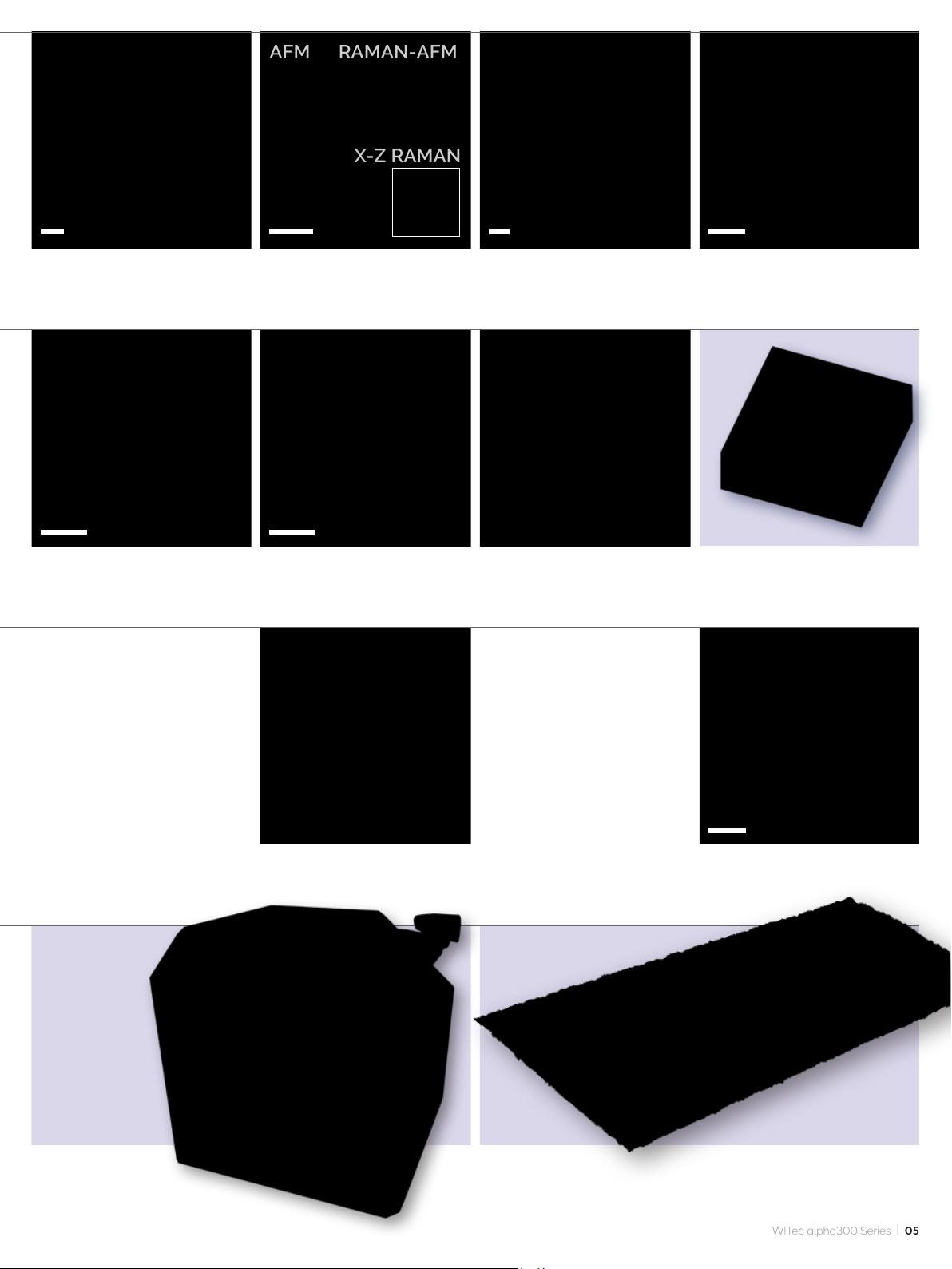
Raman image of a cell Raman image of Bacillus cereus
© A. Hermelink, Robert-Koch-Institute,
Berlin
AFM image (Pulsed Force Mode)
of fossilized bacteria
Raman depth-scan image of a
drug-eluting stent
Correlative Raman-SEM (RISE) image of a geological sample with
spectra of its molecular compounds
Large-area Raman image of the gray
and white brain matter of a hamster
brain
High-resolution SNOM image
of a latex projection pattern
AFM topography image of a
steel surface
Material stress in silicon imaged
via Raman peak-shift analysis
High-resolution AFM phase image of a
drug-eluting stent surface showing the
polymer substrate structure with the
embedded drug particles
TrueSurface™ topographic Raman
image of a pharmaceutical tablet
02
2 µm 8 µm2 µm
G-layer
S2-layer
middle lamella
1 µm 2 µm
Archeology & Arts
Super-resolution SNOM
image of the nucleus
of a rat hepatocyte
AFM image of a cell's
surface
Correlative Raman –
GFP-uorescence image
of a eukaryotic cell
Raman image of a lignied
plant cell
Raman-SEM (RISE) image of
a polished cross-section of an
abalone shell
3D Raman volume image of a
pharmaceutical emulsion
TrueSurface™ topographic Raman
image of an ancient wall painting
900 µm
TrueSurface™ topographic Raman image
of paper bers
3D Raman image of a sheet of paper
1000 µm
Anorthite
Titanite
Quartz
Fluorite
Organics
Calcite
10 µm
3D Raman image of a uid inclusion
in garnet (60 x 60 x 30 μm
3
)
Correlative Raman-SEM (RISE)
image of hematite and goethite in
several crystal orientations
Large-area Raman image of a polished rock section
with corresponding spectra
© C. Heim, Göttingen University
WITec alpha300 Series
|
03
Photovoltaics &
Semiconductors
Low-
Dimensional
Materials
5 µm
10 µm
4 µm
4 µm
10 µm
Food Science
2 µm
04
AFM
RAMAN
Polymer Science,
Thin Films &
Coatings
RAMAN
3 µm
6 µm
AFM
White-light and Raman image overlay of banana pulp Emulsion used in food production (red
and green: fatty matrix; blue: aqueous
phase; yellow: emulsier)
Correlative Raman-SEM (RISE)
image of a gallium arsenide sample
(yellow: gold substrate; red: GaAs;
blue: residues from production)
AFM and Raman images of the
same sample area on a wafer
Correlative Raman-SEM (RISE) image of a graphene
ake. The colors indicate the graphene layers and
wrinkles
Peak intensity map of MoSe
2
at 19 relative
wavenumbers (cm
-1
)
© M. O'Brien, Trinity College, Dublin
Raman image of a very thin (7.1 nm)
PMMA lm (red) on glass (blue) with
a 4.2 nm alkane contamination layer
(green) on top
Correlative Raman (right) – AFM (left) images of a three-component
polymer blend
3D confocal Raman image
of a GaN layer (60 x 15 x 20 μm
3
)
Forensics
3 µm 100 nm 10 nm
RAMAN-AFM
X-Z RAMAN
AFM
10 µm
0.7 µm 0.7 µm
Nanophotonics
4 µm
SNOM image of a surface plasmon-
polariton wave launched on a
nanostructured metal grid
Correlative Raman-AFM image and
Raman x-z cross section (inset) of a
two-component ber
AFM phase image of long chain
polymer molecules
Correlative Raman-SEM (RISE)
image of a polymer blend
Raman depth-scan image of the
inner multi-layer coating of a
beverage container
High-resolution SNOM image of a WS
2
ake (left) and corresponding
photoluminescence peak position image (right)
Correlative Raman-SEM (RISE)
image of MoS
2
akes with edges
and defects
3D Raman image of carbon nanotubes
(green) on a polymer matrix (red)
Raman image of a ngerprint
with explosive material residues
TrueSurface™ topographic
Raman image of a dextrose candy
3D Raman image
of a pollen grain (green) in
crystalline honey (50 x 50 x 50 μm
3
)
WITec alpha300 Series
|
05
剩余35页未读,继续阅读
资源评论
2301_79605092
- 粉丝: 0
- 资源: 1
上传资源 快速赚钱
- 我的内容管理 展开
- 我的资源 快来上传第一个资源
- 我的收益 登录查看自己的收益
- 我的积分 登录查看自己的积分
- 我的C币 登录后查看C币余额
- 我的收藏
- 我的下载
- 下载帮助
安全验证
文档复制为VIP权益,开通VIP直接复制
信息提交成功